 A ultrasound image of a person's body
AI-generated content may be incorrect.
A ultrasound image of a person's body
AI-generated content may be incorrect.
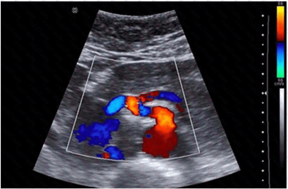
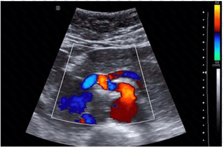
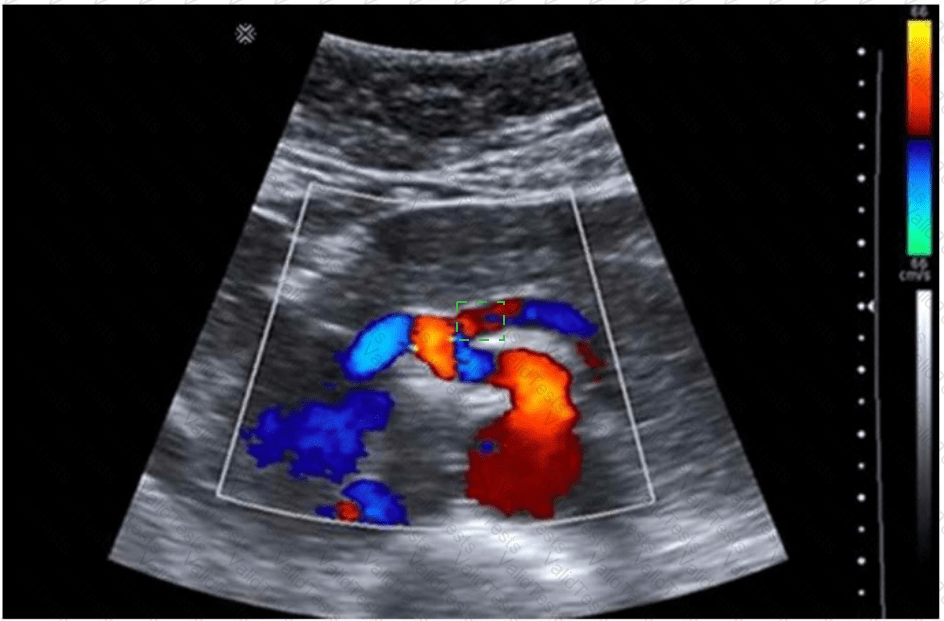
The origin of the superior mesenteric artery (SMA)
The image provided is a color Doppler ultrasound scan of the abdominal aorta and its major branches. In the center of the image, just anterior to the aorta, we see the superior mesenteric artery (SMA) arising in the sagittal plane. This is the critical area for Doppler sampling in a patient with symptoms suggestive of mesenteric ischemia.
Severe postprandial pain in a young woman may be a manifestation of median arcuate ligament syndrome (MALS) or chronic mesenteric ischemia. Both of these conditions are assessed via Doppler sampling of mesenteric vessels, specifically:
The origin and proximal segment of the SMA
The celiac artery (especially for MALS)
Doppler waveform analysis should assess:
Peak systolic velocity (PSV): >275 cm/s suggests ≥70% SMA stenosis
Angle correction should be aligned properly
Sampling must be performed at the narrowest origin point (as shown in the image)
This type of Doppler interrogation is typically done in both fasting and postprandial states to evaluate changes in flow and symptom correlation.
Why this area?
The SMA is anterior to the aorta and travels inferiorly into the mesentery.
The site shown in the image is ideal for measuring PSV and evaluating for stenosis or extrinsic compression.
[References:, Rumack CM, Wilson SR, Charboneau JW, Levine D. Diagnostic Ultrasound, 5th ed. Elsevier; 2017., Moneta GL, et al. Duplex ultrasound criteria for diagnosis of mesenteric artery stenosis. J Vasc Surg. 1991., AIUM Practice Parameter for the Performance of a Mesenteric Artery Duplex Ultrasound Examination (2020)., , , ]

Submit