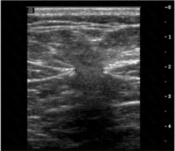
The ultrasound image demonstrates a classic example of ascites, shown by the anechoic (dark) fluid located between bowel loops or surrounding abdominal organs. In this case, there appears to be a small fluid collection in the peritoneal cavity.
One of the key maneuvers used to differentiate free fluid (such as ascites) from loculated fluid or other structures is to reposition the patient. Asking the patient to “turn from side to side” (Option D) can help in assessing whether the fluid shifts position — a hallmark feature of free intraperitoneal fluid. This positional change is highly useful in confirming the diagnosis and distinguishing ascites from other potential mimics (e.g., cystic masses, lymphoceles, or bowel wall thickening).
In contrast:
Drinking water (A) is often used in imaging the urinary bladder or gastrointestinal tract but not for fluid characterization.
Standing upright (B) may shift fluid but is less practical during real-time ultrasound.
Breathing quietly (C) doesn’t significantly aid in visualizing peritoneal fluid mobility.
[References:, Rumack CM, Wilson SR, Charboneau JW, Levine D. Diagnostic Ultrasound, 5th ed. Elsevier; 2017., Hagen-Ansert SL. Textbook of Diagnostic Sonography, 8th ed. Elsevier; 2017., AIUM Practice Parameter for the Performance of Diagnostic and Screening Ultrasound Examinations of the Abdomen and/or Retroperitoneum (2020)., , ]

Submit