 An ultrasound of a fetus
AI-generated content may be incorrect.
An ultrasound of a fetus
AI-generated content may be incorrect.
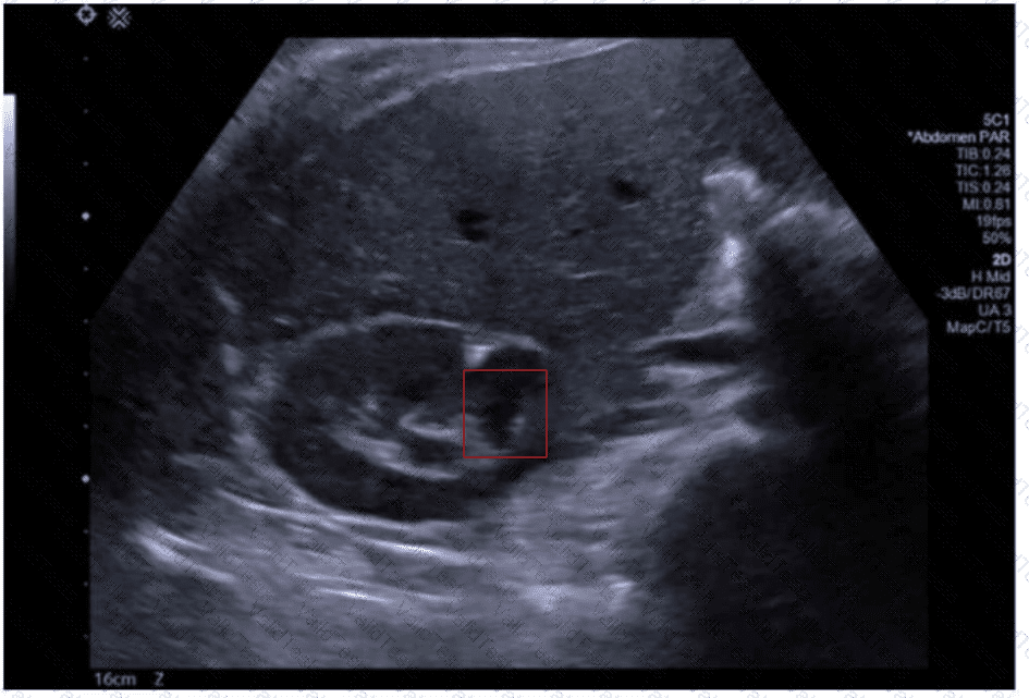
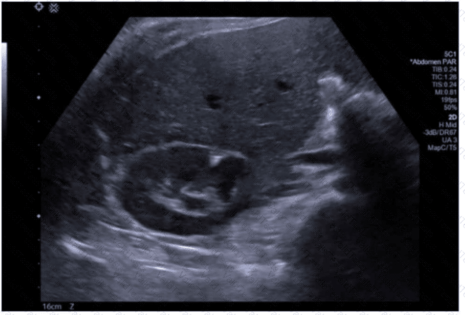
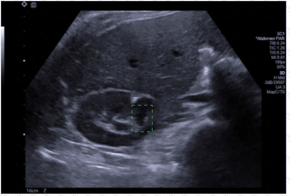
The ultrasound image shows a transverse (axial) view of the fetal abdomen. Notably, there is abnormal continuity of renal parenchyma across the midline anterior to the aorta, forming a U- or horseshoe-shaped structure. This is characteristic of a congenital anomaly known as a horseshoe kidney.
Horseshoe kidney is the most common fusion anomaly of the kidneys, occurring in approximately 1 in 400–600 live births. It results from fusion of the lower poles of both kidneys during fetal development. On prenatal ultrasound, this anomaly can be suspected when the kidneys appear closer to the midline than usual and are connected by an isthmus of renal tissue or fibrous band that crosses anterior to the spine and great vessels.
Typical sonographic findings include:
Abnormally located kidneys, often lower than expected
Renal fusion across the midline (usually at the lower poles)
Possible associated hydronephrosis or malrotation
Comparison to other anomalies:
This is not consistent with polycystic kidney disease (which would show diffusely echogenic kidneys with poor corticomedullary differentiation).
Duplex kidney would show duplicated collecting systems but not fusion across the midline.
Renal agenesis would demonstrate absence of renal tissue.
Posterior urethral valves would show a distended bladder with bilateral hydronephrosis, not midline fusion.
[References:, Rumack CM, Wilson SR, Charboneau JW, Levine D. Diagnostic Ultrasound, 5th ed. Elsevier; 2017., Callen PW. Ultrasonography in Obstetrics and Gynecology, 6th ed. Elsevier; 2016., Nyberg DA, McGahan JP, Pretorius DH, Pilu G. Diagnostic Imaging of Fetal Anomalies. Lippincott Williams & Wilkins; 2003., , , ]

Submit