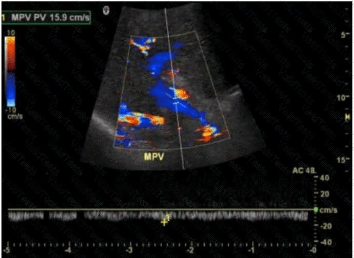
The image shows a color Doppler ultrasound of the main portal vein (MPV), which appears irregular and replaced by multiple small, serpiginous vascular channels — a hallmark of cavernous transformation. Cavernous transformation of the portal vein is a late complication of chronic portal vein thrombosis, in which collateral vessels develop around the thrombosed portal vein to bypass the obstruction.
Key Doppler ultrasound features of cavernous transformation:
Absence of a normal portal vein
Multiple tortuous vessels in the porta hepatis
Color Doppler shows hepatopetal flow in these channels
Low velocity, continuous waveform flow in collateral vessels
Differentiation from other options:
B. Portal vein thrombosis: Would show an absence of color flow within the portal vein lumen and possibly echogenic material within the vessel. There would be no serpiginous collateral vessels yet if it's an acute process.
C. Portal hypertension: Often diagnosed with other sonographic findings (e.g., splenomegaly, reversed portal flow, varices) but not characterized by the replacement of the portal vein by collateral vessels.
D. Tumor extension: Typically appears as echogenic intraluminal material within the portal vein with arterial waveforms on Doppler due to neovascularity. Tumor thrombus can be seen in hepatocellular carcinoma or pancreatic cancer, not multiple small collateral vessels.
[References:, Rumack CM, Wilson SR, Charboneau JW, Levine D. Diagnostic Ultrasound. 5th Edition. Elsevier, 2018. Chapter: Portal Venous System, pp. 107–110., American Institute of Ultrasound in Medicine (AIUM). Practice Parameter for the Performance of a Vascular Ultrasound Examination, 2021., Radiopaedia.org. Cavernous transformation of the portal vein: https://radiopaedia.org/articles/cavernous-transformation-of-the-portal-vein, , , ]

Submit