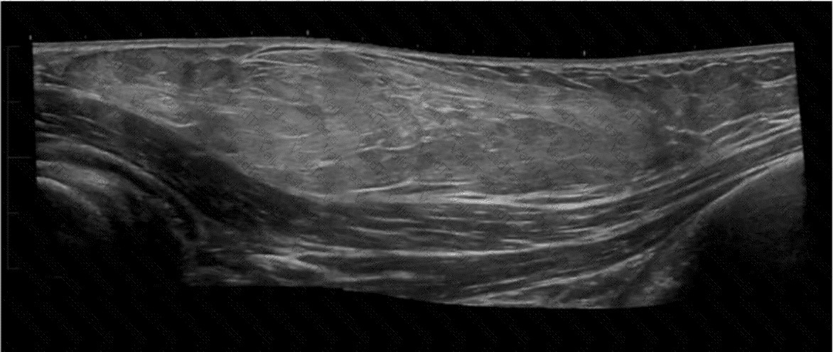
The ultrasound image shows disruption of the normal fibrillar echotexture of a muscle or tendon, consistent with a soft tissue injury such as a muscle or tendon tear. There is likely hypoechoic fluid consistent with a hematoma or edema, which commonly results from blunt or direct trauma.
This image is typical of a traumatic injury (e.g., partial or complete tendon rupture or muscle strain/tear). These findings are frequently encountered in athletic injuries or blunt force trauma and correlate strongly with the clinical history of trauma.
Key sonographic features suggestive of trauma:
Discontinuity or heterogeneity of normal striated muscle or tendon pattern
Hypoechoic or anechoic area representing hematoma or fluid collection
Retraction of muscle or tendon ends (in full-thickness tears)
Surrounding soft tissue edema
Differentiation from other options:
B. Focal pain: While pain may be a symptom, trauma is the more definitive and primary clinical indication for the findings shown.
C. Palpable abnormality: May suggest a mass or cystic lesion (e.g., lipoma, abscess), not typically the appearance shown here.
D. Decreased range of motion: May be present secondarily, but not the most consistent or primary clinical indication in this case.
[References:, Bianchi S, Martinoli C. Ultrasound of the Musculoskeletal System. Springer, 2007. Chapters on Muscle and Tendon Injuries., American Institute of Ultrasound in Medicine (AIUM) Practice Parameter for the Performance of a Musculoskeletal Ultrasound Examination, 2020., Radiopaedia.org. Muscle tear (ultrasound):https://radiopaedia.org/articles/muscle-tear-ultrasound, ]

Submit